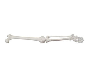
In medical education, anatomy is considered one of the most challenging basic courses. In particular, the lower limb skeleton has a complex structure, involving multiple joints, muscle attachment points, vascular and nerve channels, and it is often difficult to form an intuitive spatial concept only by learning from books and two-dimensional pictures. Therefore, the lower limb bone model has become an important auxiliary tool for medical students, which greatly improves the efficiency and understanding depth of anatomical learning.
Educational Research: How can model teaching improve Anatomy Learning?
According to research in the Journal of Anatomy Sciences Education, students who study using Anatomical models score an average of 15 to 20 percent higher on anatomy exams than those who rely solely on books. Studies have shown that anatomical models enhance spatial cognition and help students better understand the anatomical relationships between bones, especially in the lower limb regions such as the femur, tibia, fibula and their associated joints (hip, knee, ankle).

In addition, an instructional survey by the American Association for Medical Education (AAMC) found that more than 85 percent of medical students believe anatomical models are better for memorizing and understanding bone structure than books and pictures. In the clinical practice stage, students need to quickly identify the fracture site and judge the injury mechanism, and through anatomical model training in advance, they can effectively improve the ability of bone localization and pathological analysis.
Teaching Practice: How can Lower limb bone models help medical students?
1. Improved spatial cognition
- Anatomical images in books are two-dimensional, while human bones are three-dimensional structures, and it is difficult to understand the spatial relationships of bones by reading alone. The 3D three-dimensional bone model of lower limbs can help students form accurate spatial cognition and visually observe the shape, direction and articular surface of bones.
2. Improve tactile memory
- Studies have shown that Tactile Learning can deepen memory, and when using anatomical models, students can touch, move bones and simulate joint movements with their hands to more intuitively understand the interrelationships of the various parts of the bone.
3. Help clinical application
- Clinical issues such as fracture types, artificial joint replacement, and ligament injury require medical students to have an in-depth understanding of the structure and function of the lower limb bones. Using detachable or dynamic joint models, clinical surgical operations can be simulated to help students establish clinical thinking in advance.
Data support: The effectiveness of model teaching
Improved Anatomical test scores: Students using anatomical models can perform 15-20% better on anatomy tests (Anatomical Sciences Education).
- Enhanced interest in learning: Medical education research shows that more than 90% of students believe that anatomical models enhance interest in learning and make abstract anatomical knowledge more vivid (AAMC data).
- Improved clinical recognition: In imaging and orthopedic training, students using model exercises were 25% more accurate in identifying fracture locations on X-rays and CT scans (data from medical imaging studies).