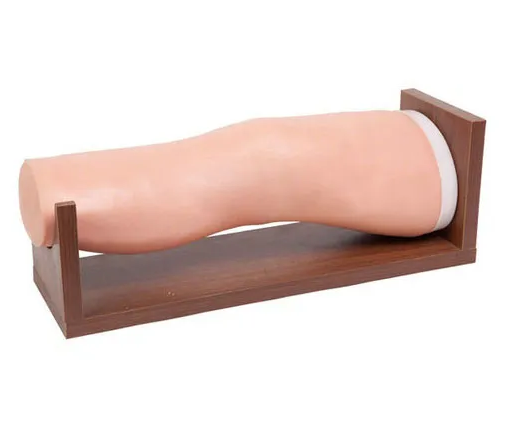
The knee cavity injection model provides a valuable tool for medical education and clinical practice with its high simulation and accuracy in simulating the process of knee joint puncture. Here is a detailed description of how the knee cavity injection model simulates knee puncture:

1. Model characteristics
The knee cavity injection model usually simulates the shape and internal structure of adult legs, especially the anatomical structure of the knee joint, such as tibia, femur, collateral ligament, cruciate ligament, patellar ligament, fat pad, meniscus and synovial sac, etc., to ensure the accuracy and authenticity of puncture training. In addition, the model is usually made of polymer material, which is easy to enter the needle and has a realistic feeling of entering the needle, and the simulated bursae has a good needle-resistant effect.
Second, the simulation process
Position preparation: The patient (model) lies supine on the operating table with both lower limbs extended to simulate the patient's position in actual clinical operation.
Disinfection and anesthesia: The skin of the puncture site should be disinfected according to the usual procedures. The physician should wear sterile gloves, lay down a disinfectant towel, and apply 2% lidocaine for local anesthesia.
Selection of puncture points: Standard knee joint puncture points are usually marked on the model, such as the junction between the outer upper margin of the patella and the lateral femoris muscle, the lower margin of the patella, and the outer 1cm of the patellar ligament. The trainer can choose the appropriate puncture point according to different situations.
Puncture operation: Use a 7-9 needle to puncture at the predetermined puncture point and Angle. For example, in the puncture method of the outer upper margin of the patella, it is necessary to press the lower depression of the lateral femoral muscle and stick the nail into 0.5-1cm; In the patellar puncture method, the knee should be bent at 90 degrees, and the 10-point needle should be parallel to the tibial plateau and punctured inward at a 45 degree Angle.
Extraction and injection: Simulate the extraction of accumulated fluid. If you need to inject drugs, you should replace the sterile syringe.
Postoperative treatment: The puncture site was covered with sterile gauze and then fixed with tape.
Third, precautions
Puncture instruments and surgical procedures should be strictly disinfected to prevent secondary infection of sterile joint cavity seepage.
The movement should be gentle to avoid damaging the joint cartilage.
If there is too much fluid in the joint cavity, appropriate pressure should be applied to fix it after suction.
The simulation training of knee joint puncture through the knee cavity injection model can effectively improve the operation skills and clinical resilience of medical personnel, and lay a solid foundation for practical clinical work.