
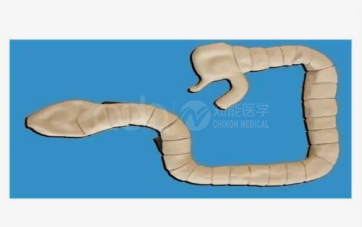
Quick answer. The BIX-LV42 is a large-intestine model mounted on a base plate, with the colon outline and intraluminal structure visible. Sigmoidoscopy on the model reveals hyperplastic polyps and cancer lesions, and multiple lesion types are provided so trainees can practise distinguishing benign from malignant change and practise intraluminal photography.
Endoscopy training has an awkward first phase: the trainee must learn to see before being allowed to steer. On a live patient, the supervisor controls the scope and the trainee watches a screen. A bench model reverses that order — the trainee looks, names, and only later handles an instrument in a patient.
| Feature | Detail | Teaching use |
|---|---|---|
| Colon outline | Large intestine form on a base plate | Orientation: where the sigmoid sits and why the approach matters |
| Lumen | Internal structure visible through the channel | Identifying folds, lumen direction and blind spots |
| Lesions | Hyperplastic polyps and cancer, multiple types supplied | Recognising and describing lesions |
| Differentiation | Benign vs malignant change | Building the vocabulary of morphology, not just detection |
| Photography | Practise intraluminal imaging technique | Documentation habits before live procedures |
A trainee who can find the lumen but cannot describe what is in it adds time and risk without adding diagnostic value. Teaching morphology first — sessile versus pedunculated, surface pattern, ulceration, friability — gives the supervised phase a shared language.
The evidence for simulation in endoscopy is strongest at the early phase. Systematic reviews of skills transfer after surgical simulation report consistent benefit for procedural skills acquired in a simulated setting before clinical exposure [1]. Reviews of virtual reality simulation in endoscopy training reach a similar conclusion, with the greatest gains among novices rather than experienced endoscopists [2]. A Cochrane review of virtual reality simulation for gastrointestinal endoscopy trainees found that simulation-based training improves early performance compared with no simulation, while noting that the effect diminishes as trainees gain clinical experience [3].
That pattern is exactly what a low-cost bench model exploits: it is a novice-stage tool. Do not buy it expecting to train advanced polypectomy.
Keep the description task written. Speaking a finding is easy; writing it forces precision.
Quotation. Lesion set contents, base dimensions and pricing for the BIX-LV42 are available from adanursing@adaanatomy.com; mention whether you need it for nursing endoscopy assistance training or for medical novices.
Q1. What can be seen inside the BIX-LV42? The colon outline and intraluminal structure, including hyperplastic polyps and cancer lesions, with multiple lesion types supplied.
Q2. Can trainees distinguish benign from malignant lesions on it? Yes. Identifying benign and malignant pathological change is one of the stated uses.
Q3. Is it a full colonoscopy simulator? No. It is a bench model for lesion recognition and orientation, not for scope navigation or loop management.
Q4. Does it support photography practice? Yes. The model allows intraluminal photography technique to be practised.
Q5. Who is it for? Novice endoscopists, endoscopy nurses and students who need to recognise lesions before supervised live procedures.
Q6. How is it stored? On its base plate, covered, away from heat. The lumen should be cleaned according to the supplier's protocol after handling.
Product page: BIX-LV42 Sigmoidoscope Simulator: Lesion Recognition First — specification, packing data and a quotation request form.
For the BIX-LV42 lesion set or a quotation, write to adanursing@adaanatomy.com.