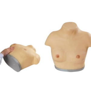
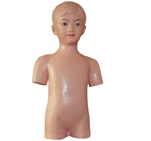
The upper and lower limb model is an important medical teaching and clinical auxiliary tool, which is widely used in medical education, clinical training, pathological analysis and rehabilitation treatment. This model is usually composed of real or simulated bones, muscles, joints and ligaments, aiming to help medical students, doctors and patients understand the anatomical structure, function and movement mechanism of the lower limbs more intuitively. With the increasing demand for 3D visualization tools in the medical field, the application of upper and lower limb models in various medical fields has become increasingly important, occupying approximately 50% of the significant position in modern medical education and clinical treatment.
1. Application in Medical Education
In medical education, the upper and lower limb models are used as teaching tools to help students comprehensively learn the structures of the lower limbs such as bones, muscles, ligaments, joints and blood vessels. Unlike traditional anatomy books or floor plans, three-dimensional models can more intuitively display various parts of the thighs, knees, calves and feet, greatly enhancing learners' understanding of lower limb anatomy. Especially in anatomy courses, students can directly operate models and observe the interaction between different parts, which is of great help in deepening memory and improving learning efficiency.
2. Clinical training and surgical simulation
The upper and lower limb models also play an irreplaceable role in clinical training. Surgeons, orthopedic doctors, rehabilitation therapists and others can become familiar with various surgical operations, treatment procedures and first aid techniques through the operation of models of the upper and lower limbs. For instance, in the simulation of fracture fixation, joint replacement surgery and knee arthroscopic surgery, the model provides a training platform with low risk and high repeatability. Doctors can improve their surgical skills and understanding of anatomical structures through continuous operation.
3. Diagnostic and therapeutic assistance
Models of the upper and lower extremities can also play a significant role in the diagnostic process, especially in the diagnosis of joint diseases, sports injuries and fractures. Doctors can infer the patient's condition by observing the joint movement, fracture type and injury site of the model, and then formulate a personalized treatment plan. For instance, when it comes to knee joint injuries, doctors can intuitively understand different types of knee problems (such as ligament ruptures, meniscus injuries, etc.) through models and accordingly select appropriate treatment methods.
4. Patient education and communication tools
The upper and lower limb models are not only tools for professional medical staff but also play a significant role in patient education. Through the intuitive display of the model, doctors can clearly explain the patient's condition, treatment methods and postoperative precautions. For instance, before undergoing knee replacement surgery, doctors can use models to explain to patients the surgical procedures, postoperative recovery precautions, and the range of motion of the joint after the operation, thereby enabling patients to have a clearer understanding and expectation of the surgical process.
The application of upper and lower limb models undoubtedly occupies approximately 50% of the significant position in the modern medical field, especially playing an indispensable role in medical education, clinical diagnosis, treatment, rehabilitation training, and patient communication. It not only enhances the technical proficiency of medical staff, but also enables patients to better understand their conditions and treatment plans during the treatment process. With the development of technology, future models of the upper and lower limbs will be more accurate and multi-functional, providing stronger support for the development of the medical industry.
...